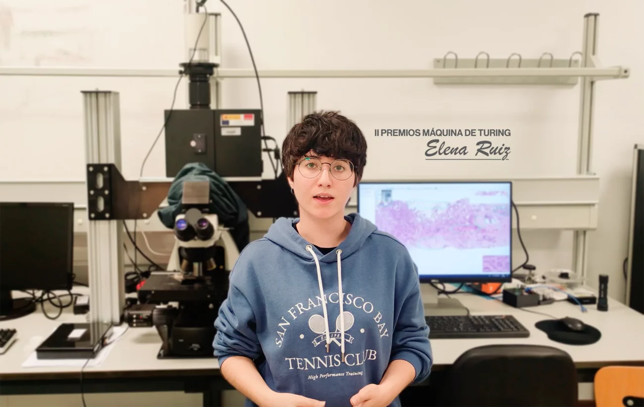
Video | Best TFG – Breast Cancer Identification
The work entitled: “Digital staining in multispectral microscopic images. Application to the identification of breast cancer”, developed by Elena María Ruiz Izquierdo and directed by Jesús Salido Tercero and María Gloria Bueno García”, was one of the End of Studies Projects (TFE) awarded in the 2nd edition of the Máquina de Turing. You can see a summary of the work through the following video:
Summary:
Artificial Intelligence is a technology that has gradually been incorporated into our lives. In particular, Computer Vision stands out for its use in various fields such as: security, automatic navigation, medicine, etc. In this way it has become one of the disciplines on the rise of the moment. The application of this technology in the field of medicine has facilitated great advances, assisting in the diagnosis and analysis of diseases, lowering costs and streamlining processes.
The purpose of this project is the classification of multispectral images of breast tissue biopsies with the help of convolutional neural networks that allow detecting tumor tissue and discriminating tumor samples from non-tumor samples. For this, multispectral images of complete histological samples of breast tissue have been used, taken in a range of 425nm to 697nm in an interval of 4nm, which makes a total of 69 wavelengths. With these samples, it is possible to identify and characterize the map of the tumor area and its different tissues. Images of unstained and biomarker-stained histology preparations or samples have been used for comparison.
The images of the samples without staining were acquired at different wavelengths with an optical microscope available with a hyperspectral camera. For this reason, an analysis was carried out to determine if they provide additional information to that obtained with images acquired with common cameras and, furthermore, if it is possible to classify these images into malignant and benign tissue. As a result, the comparison between the classification models implemented with the multispectral images is presented, to conclude which of them obtains a higher performance with a success rate closer to the classification model of images of stained preparations.
The creation of an image classifier of tissue without staining with any biomarker will eliminate the variability derived from the preparation and interpretation of the samples, also avoiding its high cost. In order to subsequently validate these techniques with the current methodology based on stained samples, several digital staining methods have been implemented applying deep learning techniques. The modeling of these data will also allow progress in the analysis of structural interactions at the cellular and tissue level.


